Cataracts: A Comprehensive Guide to Understanding, Preventing, and Managing Lens Opacity
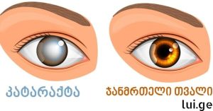
Cataracts are a condition characterized by the clouding or loss of transparency of the lens in any part of the eye. It is among the most common eye disorders globally and is a leading cause of reversible vision loss. Cataracts can affect one or both eyes and may develop gradually or suddenly, depending on their type and underlying causes. Understanding cataracts involves exploring their anatomy, etiology, symptoms, diagnostic procedures, treatment options, and preventive strategies.
1. Anatomy of the Eye and Lens Function
The eye is a highly specialized organ that converts light into neural signals, which are then interpreted by the brain to create the perception of vision. Central to this process is the lens, a transparent, flexible, biconvex structure located behind the iris and pupil. Its primary function is to focus light onto the retina, allowing for sharp and clear vision at varying distances.
The lens consists mainly of water and proteins, arranged in a precise structure to maintain transparency and refractive properties. Over time, protein aggregation and other changes may disrupt this structure, leading to opacification known as cataracts. The lens is avascular, meaning it does not have a direct blood supply; instead, it receives nutrients through the aqueous humor, a clear fluid in the anterior chamber of the eye. This avascularity contributes to the lens’s susceptibility to age-related changes and limits its natural ability to repair damage.
2. Types of Cataracts
Cataracts can be classified based on their location within the lens and the cause of opacification:
a. Nuclear Cataracts
These form in the central zone (nucleus) of the lens. They are usually age-related and progress slowly, often causing a shift toward nearsightedness (myopic shift) as the lens thickens.
b. Cortical Cataracts
These occur in the lens cortex, the outer layer surrounding the nucleus. They appear as wedge-shaped opacities that extend from the periphery toward the center, often affecting contrast sensitivity and causing glare during night driving.
c. Posterior Subcapsular Cataracts
Located at the back of the lens beneath the capsule, these cataracts are more common in individuals with diabetes, prolonged steroid use, or radiation exposure. They tend to progress faster and significantly impair near vision and reading ability.
d. Congenital Cataracts
Present at birth or developing during childhood, these may result from genetic disorders, intrauterine infections, or metabolic conditions. Early detection and treatment are crucial to prevent amblyopia and long-term visual impairment.
e. Secondary and Traumatic Cataracts
Secondary cataracts arise due to systemic diseases such as diabetes or as a side effect of medications like corticosteroids. Traumatic cataracts result from physical injury, chemical exposure, or radiation affecting the lens structure.
3. Symptoms of Cataracts
Cataracts manifest with a wide range of symptoms that may vary depending on the type and severity of lens opacity:
Gradual blurring or clouding of vision
Increased difficulty seeing at night or in dim light
Glare and halos around lights, particularly at night
Fading or yellowing of colors
Double vision in a single eye
Frequent changes in eyeglass prescription
Sensitivity to bright sunlight
As cataracts progress, patients may experience both near and distance vision impairment. The cloudiness of the lens affects the transmission and refraction of light, leading to visual distortion. Nighttime driving may become particularly challenging due to glare from oncoming headlights and streetlights.
4. Causes and Risk Factors
Cataracts develop due to a combination of aging, environmental factors, genetics, and systemic health conditions.
a. Age and Protein Degeneration
The lens grows throughout life. Protein fibers in the nucleus continue to accumulate, leading to thickening and clouding over time. This age-related process is the most common cause of cataract formation, typically noticeable after the age of 55.
b. Diabetes and Metabolic Disorders
High blood sugar levels can alter lens metabolism, causing osmotic stress and accumulation of sorbitol, contributing to cataract formation. Diabetic patients often develop cataracts earlier than non-diabetic individuals.
c. Ultraviolet (UV) Radiation
Chronic exposure to sunlight without adequate eye protection increases oxidative stress in lens proteins, accelerating cataract formation. UV-B rays are particularly damaging.
d. Medications and Lifestyle Factors
Long-term use of corticosteroids, certain diuretics, and some anti-inflammatory medications can increase cataract risk. Smoking and excessive alcohol consumption are also recognized contributors.
e. Genetic Predisposition
Family history of cataracts can increase susceptibility, particularly for congenital or early-onset cataracts.
f. Ocular Trauma and Inflammation
Injuries or chronic ocular inflammation (uveitis) can disrupt lens structure and trigger cataract formation.
5. Diagnosis
Diagnosing cataracts requires a comprehensive ophthalmologic examination:
Visual Acuity Testing: Measures clarity of vision using eye charts.
Slit-Lamp Examination: Magnifies and illuminates the anterior structures, allowing detailed inspection of lens opacities.
Retinal Examination: Evaluates the retina through dilated pupils to ensure other causes of vision loss are ruled out.
Tonometry: Measures intraocular pressure to detect coexisting glaucoma.
Contrast Sensitivity and Glare Testing: Assesses functional vision impairments not detected by standard acuity tests.
Early diagnosis is essential to prevent progressive vision loss and guide timely intervention.
6. Treatment Options
Cataract surgery is the definitive and highly effective treatment for lens opacity. Conservative measures, such as updated eyeglass prescriptions, may temporarily improve vision in early stages, but surgery is ultimately required for significant functional impairment.
a. Surgical Techniques
Phacoemulsification: The most common modern technique involves using an ultrasonic device to fragment and aspirate the clouded lens through a small incision (2–3 mm). No sutures are typically required.
Extracapsular Cataract Extraction (ECCE): Used in advanced cases, the lens nucleus is removed in one piece through a larger incision.
Laser-Assisted Cataract Surgery: Enhances precision in capsulotomy and lens fragmentation using femtosecond lasers.
b. Intraocular Lens (IOL) Implantation
After lens removal, an artificial lens is implanted:
Monofocal IOL: Provides clear vision at a single distance, typically far.
Multifocal or Extended Depth-of-Focus IOL: Allows for near and intermediate vision with reduced dependence on glasses.
Toric IOL: Corrects pre-existing astigmatism.
c. Anesthesia and Recovery
Most surgeries are performed under local anesthesia with or without mild sedation. General anesthesia is rarely needed. Patients usually return home the same day. Postoperative care includes anti-inflammatory and antibiotic eye drops, activity restrictions, and follow-up visits to monitor healing.
7. Potential Complications
Though cataract surgery is safe, rare complications may include:
Infection (endophthalmitis)
Increased intraocular pressure
Posterior capsule opacification (secondary cataract)
Retinal detachment
Corneal edema or decompensation
Appropriate surgical technique and postoperative care minimize these risks.
8. Prevention and Lifestyle Recommendations
While aging cannot be prevented, certain lifestyle modifications may reduce cataract risk or delay progression:
Wear UV-protective sunglasses
Maintain blood sugar within normal limits
Avoid smoking and limit alcohol consumption
Consume a diet rich in antioxidants (vitamins C, E, lutein, zeaxanthin)
Regular eye exams to monitor lens health
9. Nutrition and Eye Health
Specific nutrients contribute to lens protection and visual function:
Vitamin C: Protects lens proteins from oxidative damage.
Vitamin E: Antioxidant properties support lens transparency.
Lutein and Zeaxanthin: Concentrated in the lens and retina, improve light filtration and reduce oxidative stress.
Omega-3 Fatty Acids: Support retinal and lens function, may reduce age-related visual decline.
Dietary sources include leafy greens, citrus fruits, nuts, seeds, and fish.
10. Psychological Impact and Patient Support
Vision impairment affects quality of life, independence, and emotional well-being. Patients may experience:
Anxiety and depression
Reduced mobility and social engagement
Dependence on caregivers
Support includes counseling, vision rehabilitation, and peer support groups to ensure a smooth transition before and after surgery.
11. Case Studies and Patient Experiences
Case Study 1: A 62-year-old patient with nuclear cataract experienced gradual blurring and difficulty reading. After phacoemulsification with monofocal IOL implantation, visual acuity improved to 20/20, allowing return to independent living.
Case Study 2: A 55-year-old with posterior subcapsular cataract reported glare and impaired near vision. Multifocal IOL implantation restored both distance and near vision, reducing dependence on reading glasses.
Patient education on symptoms, lifestyle, and postoperative expectations is critical for successful outcomes.
12. Future Directions in Cataract Management
Emerging research explores:
Pharmacological agents to delay cataract progression
Advanced IOL materials with enhanced UV and blue-light filtration
Non-invasive imaging techniques for early detection
Gene therapy for congenital cataracts
While these innovations hold promise, surgical intervention remains the standard of care.
Conclusion
Cataracts are a prevalent and treatable cause of vision loss. Understanding the anatomy, risk factors, symptoms, diagnostic procedures, treatment options, and preventive measures empowers patients to make informed decisions and maintain quality of life. Regular eye exams, a nutrient-rich diet, UV protection, and timely surgical intervention are key components of comprehensive cataract care.
By embracing both modern medicine and lifestyle modifications, individuals can preserve vision and enjoy independence well into later life.